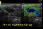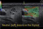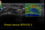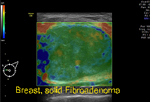
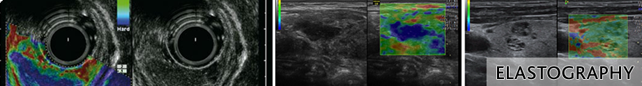
It is based on the principle - when a mechanical compression or vibration ( stress ) is applied, the tumor deforms ( strain ) less than the surrounding tissue. A tumor or a suspicious cancerous lession is normally 5 – 28 times stiffer than the back ground of normal soft tissue. Physicians have traditionally examined textural differences in tissue during physical exams with hand palpation. Elastography is the 21st century electronic interpretation of the physical exam. It is a useful adjunct to high resolution ultrasound in borderline cases for differentiating between benign and malignant lesions and reducing number of biopsies especially in the breast. Clinical applications of this feature is useful in superficial structures such as in breast, prostate, thyroid, cervical lymph nodes and evaluation of the cervix for estimating risk of pre term labor.
|
|
|||||||||||||||||||||
|